


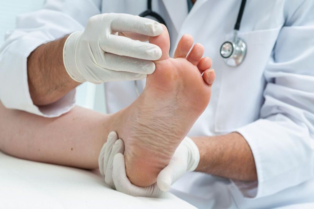
In this article, we will consider the main types of foot fungus.
All fungal infections are collectively called mycosis. Experts divide it into two main categories - onychomycosis and dermatomycosis. In the first case, microorganisms penetrate under the nail, in the second, the skin is affected. Furthermore, all types of foot fungus are classified taking into account the main causative agents of the disease and symptoms.
The main type
First of all, the type of fungus that affects the feet depends on the causative agent of the disease. Pathogenic microorganisms are divided into several groups: dermatophytes, yeasts and molds. They can provoke common injuries such as:
- Onychomycosis;
- Candidiasis;
- Epidermophytosis;
- Rubrophytia.
The latter is the common name for a group of pathologies in which the foot is affected. All diseases are also divided into several groups based on clinical manifestations: membranous, such as moccasins, vesicular.
We will consider the types of foot fungus, photos and treatments with alternative methods below.
Rubrophytia
The second name of this disease is rubromycosis. It is distinguished by congestion of blood vessels, dryness and severe detoxification. The pathology develops very slowly and is almost invisible for a person infected with the infection.
Foot fungus in the form of bubbles is very unpleasant.
The first signs are itching and flaking, which become noticeable in the later stages of the disease. Damage to the nails also occurs at this stage. Externally, the disease is manifested by the appearance of blisters, crusts, plaques, pustules, which are localized to the entire surface of the soles of the feet. When a large number of plaques and bubbles appear, a person begins to experience a painful sensation while walking.
Determining the type of foot fungus (pictured) plays an important role in treatment. Before prescribing therapy for rubrophytosis, it is necessary to perform microscopy and study the clinical picture. Lesion removal is performed using an exfoliating (keratolytic) agent. For the most part, these are ointments and creams, which are based on salicylic acid. Therapy is usually complex. In parallel with external agents, anti-fungal agents are prescribed.
If the disease is severe, you should start taking the medicine in pill form. Lesions on the nail plate are treated by removing them with an emollient.
This type of foot fungus (see picture above) is characterized by a high level of infection. Just touch on the things used by the carrier of the infection. The chances of infection increase many times over if a person suffers from excessive sweating, has a weakened immune system and damage to the legs.
The causative agent of this disease is the fungus Tr. Mentagrophytesvar. It can penetrate the granular skin layer and stratum corneum, spread and cause allergic reactions and other severe types:
- pain while walking, burning and itching;
- deform and yellowing of nails;
- appearance of crusts, scales, painful cracks;
- skin erosion (massaging);
- appearance of pustules, edema;
- vesicle rash with a dense crust.
The diagnosis of this type of foot fungus consists in the study of external signs and clinical picture. If the causative agent of the pathology is unclear, clinical research may be required, for example, examining the erosion under a microscope.
Therapy of rubrophytosis in the acute form involves the use of drugs based on silver nitrate 0. 25%, calcium 10%and meta-dihydroxybenzene 1%. If an allergic reaction occurs, antihistamines should be used. The choice of antifungal agent depends entirely on the clinical course of the disease and the individual characteristics of the patient's body.
What types of foot fungus are there?
Candidiasis of the feet
This type of fungus occurs in patients more rarely than epidermophytosis or rubromycosis. Pathology occurs under the influence of fungi of the genus Candida. Such microorganisms live in everyone's body, however, they are conditionally considered pathogenic. That is, they do not pose a threat in small numbers, but their rapid multiplication can lead to unpleasant symptoms and consequences. Uncontrolled fungal reproduction begins if there is a decrease in immunity during hypothermia, from too much work or frequent stress. External factors include:
- wear, especially in summer, uncomfortable shoes;
- injuries received at home or at work;
- persistent dryness of the skin of the feet (exfoliation of the skin due to prolonged exposure to water).
There are two types of foot candidiasis: hyperkeratotic and vesicular-pustular. The first form of candidiasis is characterized by thickening of the stratum corneum. Above it, a rather wide groove with a light brown color began to appear, which continued to peel off. For diagnostic purposes, exfoliation is performed, and further study of the particles in which the Candida fungus is found.
The viscous-pustular form of candidiasis manifests itself in the form of hyperemia (too many blood vessels), marked swelling, massaging. The affected skin area is covered with small flat -shaped pustules and blisters. After the extinction of the inflammatory process, desquamation develops. The appointment of therapy can only be done after determining the correct diagnosis. The choice of medication for this type of foot fungus with blisters is done individually. Often, systemic and local medications are indicated.
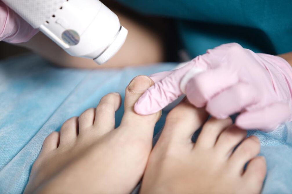
Onychomycosis
The disease is a type of fungus on the feet, which is characterized by a fungal infection on the nails. You can be infected in public baths, saunas, baths, swimming pools. Scales containing pathogenic microorganisms are easily separated from nail plates and can remain on unpainted floors, carpets, mats, and benches. High humidity allows them to not only survive, but also induce active reproduction, and therefore the risk of infection increases significantly.
In the early stages, the infection enters the epidermis of the foot, causing severe itching. To alleviate the unpleasant sensation, the person begins to comb the infected area, but the condition only gets worse. The area of skin affected by the fungus is covered with small scratches and cracks, microorganisms begin to spread, penetrating under the nail plate, after which they begin to multiply uncontrollably.
Severe diseases such as diabetes or HIV, impaired blood circulation, trauma to the nails greatly increase the risk of infection.
Onychomycosis is classified into 3 types:
- Normotrophic. With this type of onychomycosis, a change in nail color from normal to brownish-yellow is observed. The natural luster, shape and thickness of the nails remain unchanged.
- Hypertrophic. There is a final change in the color of the nail, its luster disappears, the shape changes, thickening develops and partial destruction begins.
- Onycholytic. The color of the affected nails turns brown, becomes thinner, begins to break. The gradual separation from the bed begins. In the open part of the nail bed, an uneven layer can be observed.
This type of fungal therapy on the palms and soles of the hands with topical medications is ineffective due to the fact that the fungal spores are located under the nails. Before starting treatment, the nail should be removed. This is done with keratolytic drugs, and patches are also used. In some cases, there is a possibility of mechanical nail removal: dead nail particles are cut with nails or nail polish. It is important to remember that all instruments used must be sterile.
The use of a combination of mechanical removal and keratolytic spots is the most effective method of removing diseased nails. From keratolytic agents, you can use ready -made sets, which contain special ointments, files for scraping nails, plasters. After the nail plate is removed, you should start taking systemic antimycotics.
It is quite difficult to determine the type of foot fungus from the photos.
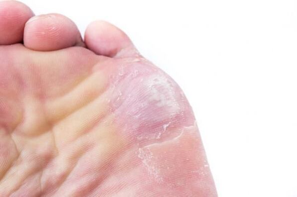
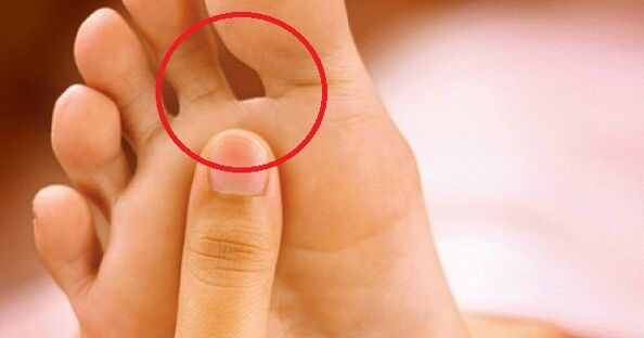
Interdigital form (intertriginous)
The most common and unpleasant type of pathology is an interrelated form of fungal infection. Appears quite often in the summer, begins to grow between the third and fourth toes. Over time, the lesion spreads to the area between the other fingers.
Initially, cracks, funnels or small sores appear in the folds located between the fingers. It is surrounded by a diaper rash or flaky skin that is slightly greenish in color. Often, the damage becomes wet, sometimes pus comes out of it. The type of fungus that is removed is marked by a clear or flour-like exfoliation, as if there is flour on the surface of the finger. A similar effect arises because of the large number of affected scales that separate from the skin. There is a slight itching that does not cause severe discomfort.
With more advanced forms of the disease, there is nail melting, severe melting, double cracks, corn -like corneal compaction, obvious yellowing.
In very rare cases, the disease of the weeping type develops - exudative fungus. The main difference is that the vesicles are poured into the affected area - bubbles filled with fluid in them. Therapy should be carried out thoroughly. Antifungal agents are used as topical agents. Advanced forms of the disease involve the use of systemic antimycotics. The course of treatment should be continued until the fungus is completely gone.
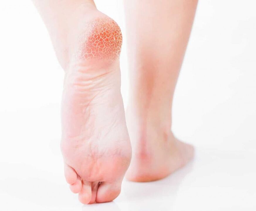
Squamous-hyperkeratosis form
This type of foot fungus (pictured below) is not very common.
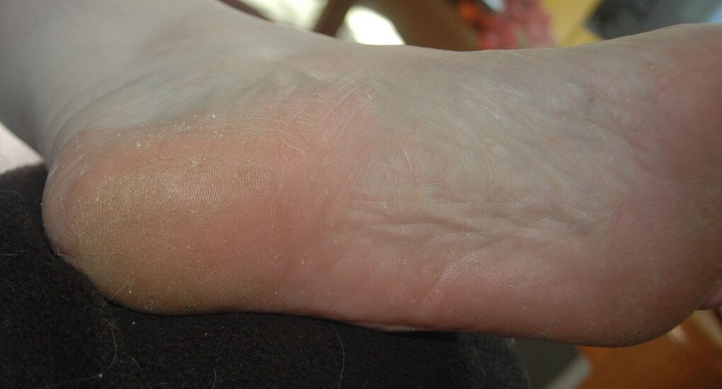
Squamosis is the process of penetration of pathogenic fungi into the outer skin cells. Hyperkeratosis is the formation of the stratum corneum, which results in thickening of the dermis. In this case, the squamous-hyperkeratotic form of mycosis has several more names, for example, "moccasin fungus" and "athlete’s foot".
Mycosis of the squamous-hyperkeratotic type is characterized by the following symptoms:
- The soles of the feet are covered with a thickened layer of keratin from the dermis, which produces the effect that moccasins are worn on the feet.
- The contraction of the palm occurs so strongly that it begins to cover with a wide and rather thick callus.
- Painful cracks appear in the corn.
- Peeling takes on a type of mucus, the pattern on the skin can be seen with the naked eye.
- Unbearable itching appears.
- Over time, the nails begin to thin, break and shatter.
When treating moccasin fungus, first of all, it is very important to remove the stratum corneum on the skin. This is done using a soap-soda foot bath, a bandage, salicylic compresses, and ichthyol ointment. Salicylic ointment is used in doses up to 10%. Duct -based creams, ointments containing lactic acid are effective. If you are unable to cope with this task at home, you should seek help from a podiatric center. With the help of a hardware manicure, the specialist will carefully remove the keratin dermis.
Subsequent treatment for the type of foot fungus depends on the type of pathogen. It should be started only after an accurate diagnosis has been made. It is not recommended to treat moccasin mycosis without first removing the stratum corneum on the skin - the active component in the composition of the drug will not be able to penetrate it and reach the focus of infection. As a result, all attempts will be canceled.
Photos of this type of foot fungus cannot describe all the unpleasant symptoms a person experiences.
Forms of menstruation
Vesicular fungus, or, as it is also called, dyshidratic mycosis is the rarest type of disease. Its main manifestation is that many vesicles fuse into conglomerates. Vesicles are vesicles that are filled with pus or nutrient fluid from the inside. When the turbidity of the fluid begins, the vesicles rupture, the boil remains in place. They begin to merge into one line, forming a clear scar on the skin. This is due to the drying and exfoliation of the skin layer.
About 70% of infections with vesicular fungi are accompanied by an allergic rash. Various bacteria and viruses begin to penetrate the boil. As a result, the disease becomes mixed, and early pathogen identification becomes more difficult. Therefore, you should see a doctor immediately as soon as the main symptoms appear (pictured): he can quickly identify the type of foot fungus and begin therapy.
And this must be done immediately. First of all, before using antimycotic drugs, the acute process should be eliminated. It is better to leave this task to a specialist: he will be able to penetrate the vesicles gently, treat the remaining ulcers with 2% boric acid and smear with a brilliant green or methylene blue solution.
Treatment of the disease in its neglected form involves the use of corticosteroid ointments. After the elimination of inflammatory processes, it is recommended to use local antimycotic agents. This will suppress the causative agent of the disease.
We continue to consider the name and type of foot fungus.
Shape deleted
Mycosis of the erased form is almost invisible, the symptoms are minimal. These include: mild itching, burning, exfoliation of the mucus type, microcracks located in the interdigital zone. If you do not see a specialist when the first signs of the disease appear, the pathology can turn into a form of onychomycosis, which is much more difficult to treat. In this case, the missed nail will grow back from one month to six.
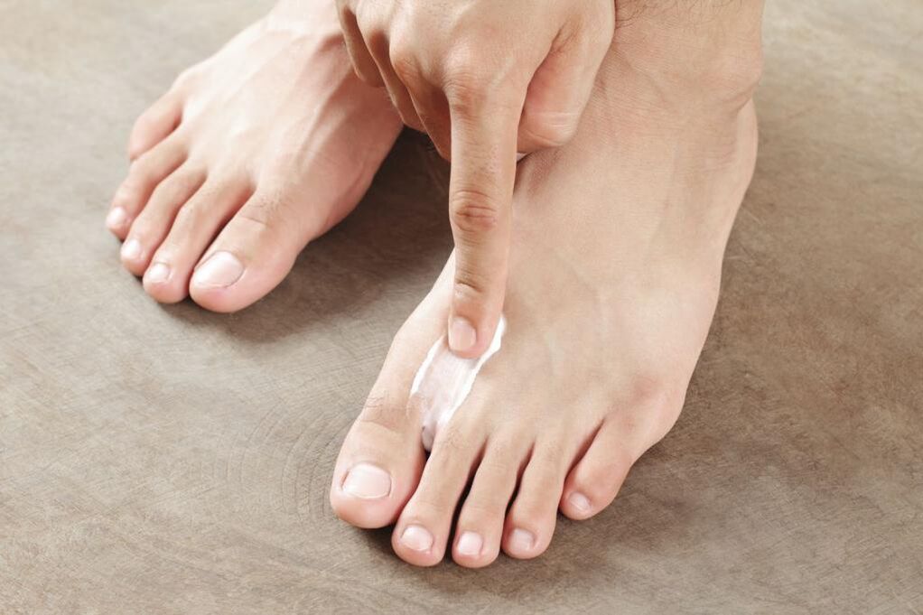
Treat mycosis of the erased form with local preparations: ointments, creams, foams. They allow you to create layers on the feet that will protect against other infections. It is not recommended to wash the feet within 24 hours after using such a drug.
Only in extreme cases, systemic therapy can be prescribed. The problem is that such drugs are toxic and negatively affect some internal organs, for example, the liver. Therefore, if there are side effects from the use of local remedies, then it is better not to take pills.
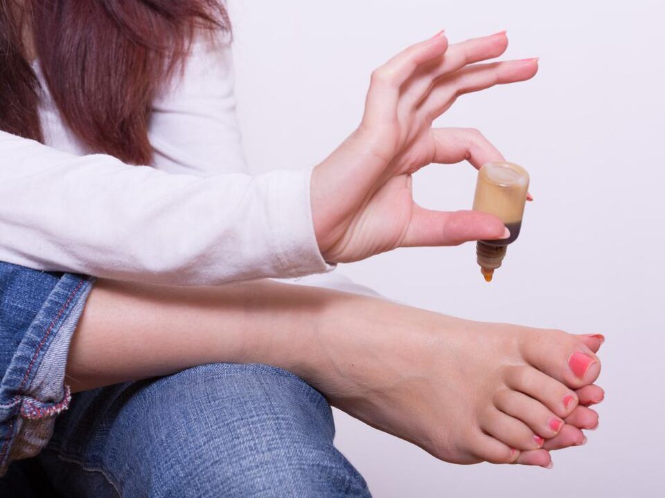
Treatment of foot fungus with alternative methods
The pathological photographs presented in the article in large numbers do not cancel the trip to the doctor.
It's easy enough to choose a drug now. However, many people prefer to treat the fungus with folk remedies. We offer several proven recipes:
- Clean the feet. The feet are heated in a basin of hot water, rubbed with laundry soap and treated with a stiff foot comb for five minutes. The foam is rinsed. The action is repeated 4-5 times. Then the feet are wiped dry and wrapped in cream.
- Celandine bath. 50 g of the ingredient is poured with 1. 5 liters of boiling water, heated on fire for 4-5 minutes, cooled. The legs should be kept in a warm broth for 30 minutes. The duration of treatment is 14 days.
- Tea tree oil is a powerful antiseptic. It must be applied repeatedly to the affected area.
- You can get rid of itching and cracks with sour cream. She lubricated her feet before bed. The duration of therapy is 1 week.
- Baking powder. Eliminates burning and itching on the skin. The powder is mixed with water to obtain a thick mass. It is applied to the affected area, wait for it to dry, then rinse.
- Calendula. Flowers (50 gr. ) Poured with boiling water (1: 2), insist 30 minutes, filtered. The infusion is lubricated feet at night.

prevention
The simplest preventative measures will reduce the chances of infection. Only personal items should be used, nails should be treated with sterile tools. When visiting public places such as baths, saunas, swimming pools, beaches, you should use your own shoes. By the way, try to choose it so that it is comfortable and allows your feet to breathe.
You should worry about prevention first, so that later you don’t have to deal with various types of foot fungus. Photos show far from all options for the development of this disease.